English

Paperback
₹6134
₹6846
10.4% OFF
(All inclusive*)
Delivery Options
Please enter pincode to check delivery time.
*COD & Shipping Charges may apply on certain items.
Review final details at checkout.
Looking to place a bulk order? SUBMIT DETAILS
Delivery Options
Please enter pincode to check delivery time.
*COD & Shipping Charges may apply on certain items.
Review final details at checkout.
About The Book
Description
Author
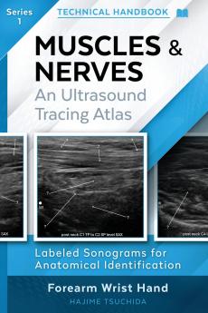
<p><span style=color: rgba(0 0 0 1)>* Master Musculoskeletal Ultrasound Anatomy: Delve into detailed ultrasound anatomy of muscles and peripheral nerves.</span></p><p><span style=color: rgba(0 0 0 1)>* Practical Scan Techniques: Gain technical explanations on how to acquire specific ultrasound structures.</span></p><p><span style=color: rgba(0 0 0 1)>* Step-by-Step Guidance: Follow along with clear step-by-step tracing of anatomical structures.</span></p><p><span style=color: rgba(0 0 0 1)>* Labeled Ultrasound Images: Easily identify structures with precisely labeled ultrasound images.</span></p><p><span style=color: rgba(0 0 0 1)>* Focused Visual Learning: Benefit from a unique one page - one image format for clear concise understanding.</span></p><p><span style=color: rgba(0 0 0 1)>* Your Essential Practical Handbook: Designed exclusively for hands-on technical application.</span></p><p></p><p><span style=color: rgba(0 0 0 1)>A practical visual atlas for clinicians performing musculoskeletal ultrasound and nerve tracing.</span></p><p></p><p><span style=color: rgba(0 0 0 1)>A Visual Step-by-Step Ultrasound Atlas for the Forearm Wrist and Hand.</span></p><p></p><p><span style=color: rgba(0 0 0 1)>MUSCLES &amp; NERVES: An Ultrasound Tracing Atlas (Series 1) is a focused practical reference designed to support clinicians and learners in recognizing and understanding musculoskeletal and peripheral nerve anatomy in real-time ultrasound imaging. This atlas provides a clear visual pathway for understanding sonoanatomy through labeled ultrasound images and anatomical tracings.</span></p><p></p><p><span style=color: rgba(0 0 0 1)>Whether you are performing MSK ultrasound guiding nerve blocks planning rehabilitation or improving diagnostic understanding this handbook supports clinical practice through clear anatomical reference-not as a substitute for medical diagnosis or treatment.</span></p><p></p><p><span style=color: rgba(0 0 0 1)>WHAT YOU WILL LEARN:</span></p><p><span style=color: rgba(0 0 0 1)>• How to confidently identify peripheral nerves and muscle layers on ultrasound</span></p><p><span style=color: rgba(0 0 0 1)>• How to follow each structure in continuity using step-by-step tracing guidance</span></p><p><span style=color: rgba(0 0 0 1)>• How to differentiate muscles fascial compartments and branching nerve patterns</span></p><p><span style=color: rgba(0 0 0 1)>• How to apply practical probe positioning techniques to achieve optimized images</span></p><p></p><p><span style=color: rgba(0 0 0 1)>WHY THIS ATLAS IS UNIQUE:</span></p><p><span style=color: rgba(0 0 0 1)>• One Page - One Image layout allows rapid comprehension during live scanning</span></p><p><span style=color: rgba(0 0 0 1)>• Each ultrasound image is paired with a precise anatomical tracing for instant orientation</span></p><p><span style=color: rgba(0 0 0 1)>• Structured for real-world scanning workflows not textbook theory</span></p><p></p><p><span style=color: rgba(0 0 0 1)>WHO THIS BOOK IS FOR:</span></p><p><span style=color: rgba(0 0 0 1)>• Sonographers and MSK imaging clinicians</span></p><p><span style=color: rgba(0 0 0 1)>• Physical and Occupational Therapists</span></p><p><span style=color: rgba(0 0 0 1)>• Orthopedics providers</span></p><p><span style=color: rgba(0 0 0 1)>• Pain medicine and regional anesthesia clinicians</span></p><p><span style=color: rgba(0 0 0 1)>• Students developing foundational sonoanatomy skills</span></p><p><span style=color: rgba(0 0 0 1)>A practical visual and clinically focused guide to mastering ultrasound anatomy of the forearm wrist and hand.</span></p><p></p>

Piracy-free

Assured Quality

Secure Transactions

Fast Delivery

Sustainably Printed
Details
ISBN 13
9781764437707
Publication Date
-02-12-2025
Pages
-112
Weight
-163 grams
Dimensions
-152x229x7.39 mm
